Wednesday, January 27, 2016

Unit 5 Reflection

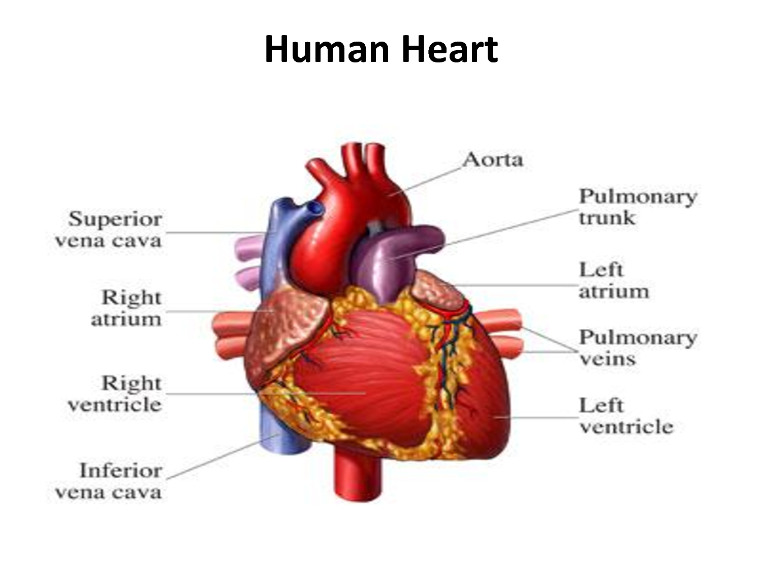
In this unit, we learned about diabetes, the endocrine system, the digestive system, and the lymphatic system. Instead of learning mostly about the anatomy and physiology of the structures in these systems, we learned about how the structures in these systems interact with other systems in the body and with different kinds of hormones in the body. One of the main themes of this unit was hormones, and how changes in levels of hormones can affect the body's systems. After we learned about the basic anatomy and physiology of the digestive system, we learned about what the body does with the nutrients absorbed from food and how hormones regulate how these nutrients are stored through the Fed State, Fasting State, and Starving State. When learning about fuel metabolism, we primarily focused on insulin, glucagon, adrenaline/noradrenaline, and cortisol. Then, we proceeded to learn about diabetes and its causes, and how it is related to the malfunction of insulin production.; we learned about the differences between Type 1 and Type 2 diabetes. Type 1 diabetes is mostly genetic and is caused by a complete inability to produce insulin. On the other hand, Type 2 is not genetic and is induced by insulin resistance. Furthermore, we learned about the endocrine system and and how hormones were secreted or stopped through a chain reaction through many glands but starting at the brain. Lastly, we learned about the basic anatomy and physiology of the lymphatic system. The lymphatic system is somewhat like a separate circulatory system that helps to absorb lipids and aids immunity.
I thought that this unit was one of the hardest units that we had ever learned, as it required lots of detail and the learning of new terms, especially for the different kinds of hormones and the glands that they came from. There were so many new terms to learn and many of the glands and hormones sounded similar to each other, so I easily confused the terms. However, this task was made easier after watching this video on YouTube:  it is a crash-course video on hormones in the human body. It was also difficult to connect the themes we learned in this unit to other units, since I usually forget some of the material learned in the previous few months. However, I felt that learning about the different fuel metabolic states was relatively easier because we also received a reading packet that made the content easier to understand.

For the fuel metabolism subject, I read an article called "Stress, Metabolism, and Liquidating Your Assets", that went into detail about what the hormones specifically do to the body and how stress and change the hormonal balance in the body. I also conducted a Digestive System Lab (http://jhan496.blogspot.com/2016/01/digestive-system-lab.html) , for which we measured the approximate lengths of our digestive systems.
Relating to this unit, I want to learn more about how genetics affect one's hormonal and digestive health, as I have read articles stating that genetics can determine the type of gut bacteria a person has; I want to know the extent to which genetics control one's hormonal control, and how much can be controlled by environmental factors.
From my New Years Goals (http://jhan496.blogspot.com/2016/01/new-years-goals.html), I feel like I have been improving in my effort to not slack off in this class. Even though I do have classes with a harder workload, I have been making sure to turn in everything despite absences in order to maintain a clean gradebook. I have also been making progress on my cello piece, and I am planning to maybe play this piece in another competition in late spring.

Tuesday, January 5, 2016

Digestive System Lab

1. In this lab, I measured the approximate lengths of the major components of my digestive system: mouth esophagus, stomach, small intestine, and large intestine. I used colored ribbon for all components except for the small intestine, and instead used regular yarn for the small intestine. From this lab, I learned that the small intestine is the longest component of the digestive system by far, since it is approximately my height multiplied by four! My small intestine measured around 660 cm, which is very long; I was surprised that all of that could fit inside of my intestinal cavity. Below is my data table from the lab:


Digestive Organ
Color and Length (cm)
Mouth
red, 11.7cm
Esophagus
blue, 45.7cm
Stomach
pink, 19.0cm
Small Intestine
yarn, 660.4cm
Large Intestine
green, 165.1cm
TOTAL
901.9cm

2. The length, in meters, of my digestive system is approximately 9.02 meters, but my height is only about 1.65 meters; the length of my digestive system is significantly longer than my height. I think that my digestive system is able to fit in my abdomen because the small and larger intestines are coiled on top of each other, so they are somewhat squeezed in a tight space within my abdomen. 

3. On average, I think that it takes about 4-6 hours for food to move through the entire digestive system. After looking it up just now, it takes approximately 6-8 hours for food to move through the entire digestive system. My guess was significantly shorter than the actual time it takes to digest food. The time it takes to digest food is influenced by the the concentration of the the food eaten; for example, whether the food is made mostly of fat, or just carbohydrates, etc. Furthermore, the amount of exercise and the level of metabolism of an individual can also influence the time it takes to digest food.

4. Digestion is different from absorption because digestion is the act of actually breaking the food down into simpler forms, so that it is possible for the body to absorb nutrients from the food. The main organs that are involved in digestion are the mouth and the saliva in the mouth, the stomach, and the duodenum of the small intestine. On the other hand, absorption is the act of absorbing the nutrients from the food, and no further breakdown of the food occurs. The main organs involved in absorption are the jejunum and ileum of the small intestine and the large intestine.

5. I want to learn the reason why the small intestine is so much longer than the large intestine, and also why the larger intestine is much wider than the small intestine. Furthermore, I also want to learn how ulcers are assuaged once they appear in the stomach wall. 

Monday, January 4, 2016

New Year's Goals

For this semester, I will pay more attention to this class and not slack off on this class's schoolwork. Because Anatomy is not my hardest class, it was difficult spending lots of time on my other harder classes and leaving little time for this class. As a result of my slacking off, my grades gradually decreased. In order to reach my first goal, I will do the following:
1. Complete my homework on time.
2. Complete all of my classwork in the time given during class to do so.
3. Make a comprehensive study guide for each test.
4. Do not skip studying for any chapters, despite the feeling that the test may be easy.

For this semester, I will also play my current cello piece, Elgar in E minor, with full confidence and master. I have been working on this piece since the beginning of the school year, and I can mostly play the piece well. However, there are still some parts of the piece that are very technically difficult, and I cannot shift fast enough to those positions. In order to reach my second goal, I will do the following:
1. Prepare for my shifts earlier to prevent delay in the first note after the shift (especially in cadenza).
2. Track all of the tenuto markings and notice the differences between tenutos on different notes (how they change the mood of the piece).
3. Establish a mood for each change in phrasing. 

Saturday, November 14, 2015

Sheep Heart Dissection

Q1: What is the purpose of the pericardium?
- The pericardium is the sac outside the heart that connects the heart to the body and protects the heart; it secretes a fluid to lubricate the heart for movement.
Q2: Observe the blood vessels connecting to the heart. How do arteries differ from veins in their structure?
- Arteries differ from veins because they have thicker walls and do not have skeletal muscles to aid the process of "milking", which veins do. Veins are surrounded by skeletal muscles that help push blood through the veins against gravity. Furthermore, arteries have elastic and contractile layers in their walls; the elastic layer expands when the heart contracts and recoils when the heart relaxes, and the contractile layer is smooth muscle that causes vasodilation or vasoconstriction.
Q3: Place your finger inside the auricle. What function do you think the auricle serves?
- The auricles are wrinkled pouches on each atrium, and they each slightly increase the amount of blood that each atrium can hold.
Q4: Observe the external structures of the atria and ventricles. What differences do you observe?
- The ventricles are significantly larger than the atria, most likely because they have to pump blood to arteries that go to the rest of the body, so there needs to be space for a larger volume of blood.
Q6: Draw a picture of the tricuspid valve, including chordae tendinae and the papillary muscle.

Q7: Why is the "anchoring" of the heart valves by the chordate tendinae and the papillary muscle important to heart function?
- The "anchoring" of the heart valves by the chordae tendinae and the papillary muscle is important to heart function because these two structures help the valves function and open to allow blood to flow in from the atrium to the ventricles. If the chordae tendinae and the papillary muscle were not attached to the heart, the valves would not be able to function properly.
Q8: Using pictures and/or words, describe what you see.
- I see the bicuspid valve in between the right atrium and right ventricle, which were smaller than I expected. There are two clear flaps/cusps with the chordae tendinae attached to them, which are also attached to papillary muscles that stick out from the walls of the chambers of the heart.
Q9: What is the function of the semi-lunar valves?
- The semi-lunar valves prevent arterial blood from re-entering the heart, so it continues to be pumped around the body.
Q10: Valvular heart disease is when one of more heart valves does not work properly. Improperly functioning heart valves can lead to regurgitation, which is the backflow of blood through a leaky valve. Ultimately this can lead to congestive heart failure, a condition that can be life threatening.
a. If the valve disease occurs on the right side of the heart, it results in swelling in the feet and ankles. Why might this happen?
- This might happen because a valve malfunction may cause blood to back up in other parts of the body. This may results in fluid buildup and cause swollen hands, wrists, feet, and ankles.
b. If the valve disease occurs on the left side of the heart, what complications would you expect to see?
- I would expect to see complications in the lungs, because the left side of the heart deals with the blood that is delivered to the lungs; there may be fluid buildup in the lungs.
Q11: Using pictures and/or words describe what you see.
- I see the bicuspid valve, which leads into the right ventricle. Then, the right ventricle curves up and I see the aortic semilunar valve, which has 3 cusps and leads into the aorta. I also see the coronary arteries on the anterior surface of the heart, since they are the main arteries that supply blood to the heart itself.
Q12: Describe how the left and right sides of the heart differ from each other.
- The left and the right sides of the heart differ from each other because the left side of the heart deals with oxygenated blood and the right side of the heart deals with deoxygenated blood. Furthermore, the left side also delivers blood through the aorta to the rest of the body, and the right side delivers blood through the pulmonary trunk to the lungs.
Q13: Draw and label all structures visible in the interior of the cross-section.
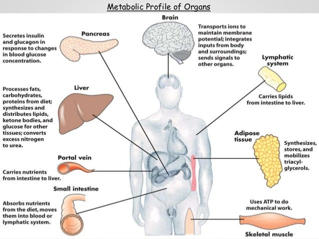
Link to our sheep heart dissection: https://www.youtube.com/watch?v=D5BNrrg4xrM


Wednesday, November 11, 2015

Monday Wellness Reflection: Guided Imagery

This Monday, my friend, Eric Sze, and I presented a beneficial health trend called guided imagery to our class. We chose this topic because our teacher had recently introduced a concept called “mindfulness” to us. Mindfulness is the phenomenon in which a person is aware of their every move and breath, essentially a form of meditation. Furthermore, at the start of class every day, the entire class participates in a short form of meditation by listening to a bell ring, closing their eyes, and breathing deeply until the ring can no longer be heard. The most interesting things that I learned while preparing for this presentation were that it actually increased certain white blood cell counts and is very commonly used in hospitals and therapy centers all over the world. When I was researching the physical benefits of guided imagery, I discovered that guided imagery increased levels of T-cells and natural killer cells, both types of white blood cells, for breast cancer patients. I also learned that many hospitals and therapy centers use guided imagery to help their patients cope with pain and treatment that they are receiving. My topic is important for health and wellness because it can be used so widely by anyone at anytime. The way someone experience guided imagery can be changed depending on how the person prefers it, which means that it can be altered depending on the person’s problem/condition. Because of that, it can be used to help cancer patients deal with pain and treatment, but can also be used on a daily basis to reduce stress and pressures of daily life. It can also be used to improve performance, whether it be mental or physical; it can improve memory and mental agility, but it can also improve sports performances. For example, famous Olympic swimmer Michael Phelps is known for thinking of a calm and relaxing place and taking deep breaths before competing in a big race. On a scale of 1 to 10, I would give myself a 9 because I think I contributed a fair amount of work into the powerpoint presentation and tried to involve the class in the activity. However, I feel as if I could have contributed more to the powerpoint in terms of content; I contributed mainly to the content of the activity and the anatomy of guided imagery, but my partner contributed the transition slides and technological aspects of the presentation, such as the insertion of video links and sources. I think that guided imagery is a very important form of meditation that should be weaved into a person’s daily schedule due to its effectiveness. Guided imagery can take as long as one wants, whether it be one minute or two hours. However, although guided imagery can be made short, reducing duration does not reduce the effectiveness of guided imagery. In the documentary about mindfulness that we watched during class, I learned that even big corporations like Google use meditation and variations of guided imagery every morning before everyone starts their work; the Google executives truly believe that these few minutes of meditation improve their performance throughout the day. Overall, guided imagery is a great technique that everyone should try to experience at least once in their lifetime!

Powerpoint: https://docs.google.com/presentation/d/1JMyUBnP6vkWn9Sko1xTnXCH3X8GZRDSxJ-ZAYUEkQeA/edit?usp=sharing

Unit 3 Reflection

Unit 3 of Anatomy and Physiology was all about the circulatory system and the different diseases that can affect the circulatory system; this included all of the main components of the circulatory system and its connections to other organ systems in the body. The cardiovascular system is made up of the heart, arteries, veins, and most importantly, blood. The heart is the body's battery that circulates blood around the body and delivers nutrients and oxygen to the tissues in the body.

Furthermore, the circulatory system also disposes of waste material from cells. The heart receives oxygen-rich blood from the lungs and is distributed to the rest of the body; the heart also receives oxygen-poor blood from the rest of the body and brings it to the lungs to dispose of carbon dioxide and other wastes collected. Cardiovascular health is the well-being of the functions and organ of the circulatory system, making sure that there is no disease present and that the person maintains a healthy lifestyle. However, many people in the world suffer from cardiovascular diseases, such as heart disease and stroke. Heart disease, which comes in many kinds (heart attack, aneurysm, atherosclerosis, etc.), is mostly caused by an unhealthy diet, lack of exercise, family history of heart disease, and unhealthy habits like smoking and drinking.

Stroke occurs when atherosclerosis or an aneurysm are present in the brain, cutting off circulation to a certain area of the brain. Since atherosclerosis and aneurysms are also considered as heart diseases, stroke is also caused by many of the factors that cause heart disease. In order to promote cardiovascular health, one should maintain a healthy lifestyle by controlling blood pressure and cholesterol, eating a healthy diet, exercising regularly, and managing stress. After finishing this unit, I still want to learn more about how HDL cholesterol clears up LDL cholesterol from clogging up the arteries. I also want to learn about the public's common misconceptions of atherosclerosis and other forms of heart disease, so I can inform people about them in order to promote cardiovascular health.

In this unit, I had trouble remembering the path of blood throughout the heart and the body; specifically, the fact that the pulmonary artery contains oxygen-poor blood confused me, since arteries typically carry oxygen-rich blood. However, I thought that I was more engaged in this unit since learning about all of the different types of cardiovascular diseases fascinated me; learning how to prevent them will also be beneficial in the future. Because of that, I thought that I retained the most information about the different cardiovascular diseases and was the most strong in that aspect. From the labs and activities from this unit, I learned that it is very hard to work on a collaborative project unless you assign each person in the group to a specific job/responsibility. For example, when we dissected the sheep heart for a lab, each person was assigned a responsibility, so the process went along quite smoothly.

My Unit 2 health goals were mainly to reduce my intake of carbohydrates and pearl milk tea and sleep at a more regular time every day. Personally, I think I have actually increased my intake of carbohydrates and pearl milk tea due to the heightened levels of stress from schoolwork and extracurricular activities. However, I do think that I have slept at a more regular time since I set my health goals for last unit. For the remainder of the semester, a few goals I have are to stay on top of my schoolwork without being overwhelmed by stress, increasing my intake of fiber/vegetables, and getting more exercising by going outside more.

Tuesday, October 13, 2015

Blood Pressure Lab

In this lab, a partner and I measured each other's blood pressure and pulse. Using a stethoscope and a sphygmomanometer, we first measured each other's systolic blood pressure.
The systolic blood pressure was measured by pumping the air bulb until the pressure reached about 150 mmHg, and then slowly releasing the pressure until a "whoosh" sound from the blood was heard. When we heard the "whoosh", we recorded the pressure (systolic). On the other hand, in order to measure the diastolic pressure, we listen through the stethoscope until the "whoosh" sound fades away; this point is called the diastolic pressure.
The heart rate is measured by a stethoscope, and the blood pressure is measured by a sphygmomanometer, a device with an air bulb, arm cuff, and a pressure reader.
Using your thumb to measure pulse is not ideal because the thumb has a pulse of its own, so it could confuse the pulse reader and make the findings inaccurate.
A sphygomomanometer requires careful and meticulous work in order to record an accurate blood pressure. First, make sure the air bladder of the cuff is deflated before wrapping it around anyone's arm. Next, wrap i around the upper arm so it fits well and make sure the velcro sticks well to ensure the cuff's position. Then, place the head of the stethoscope right under the edge of the cuff, just a small distance above the elbow crease. Inflate the cuff with quick pumps of the air bulb until the pressure reads about 150 mmHg or until the the pulse cannot be heard. Once the pressure reaches about 150 mmHg, slightly open the air flow and release some air very slowly, ensuring that the air is not released too suddenly. At this point, listen closely for any sign of blood flow while watching the pressure gauge fall. Once you hear a "whoosh" of blood flow, look at the pressure gauge and record this pressure as the systolic blood pressure. Next, watch the stethoscope and listen to the pulse until the "whoosh" sound disappears; at this point, look at the pressure gauge and record this number as the diastolic blood pressure.

Data:


Test 1
Test 2
Average for All Subjects
Pulse Rate (radial)
60 bpm
68 bpm
64 bpm
Pulse Rate (carotid)
74 bpm
66 bpm
70 bpm
Stethoscope
66 bpm
70 bpm
68 bpm
Average for Individual Subjects
66.7 bpm
68 bpm
67.3 bpm


Subject 1
Subject 2
Blood Pressure (test 1)
120/70
110/74
Blood Pressure (test 2)
122/76
107/80